Thursday, June 26, 2008

Scientists Seek To Unwrap The Sweet Mystery Of The Sugar Coat On Bacteria

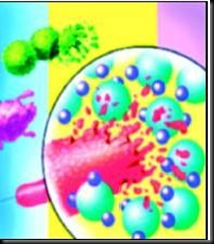
The sugars coating bacteria can change very quickly during the course of an infection, cloaking the bacteria from the immune system of their host. Previous techniques for studying the sugars were too slow to catch these rapid changes.

"There's a growing recognition of the importance of carbohydrates on bacterial cell surfaces," says Dr. Lara Mahal, lead researcher and assistant professor of chemistry and biochemistry with the Institute for Cellular and Molecular Biology. "The carbohydrate coating is critical in how your immune system recognizes bacteria."

Mahal and graduate student Ken Hsu report their findings in the advance on-line edition and March issue of Nature Chemical Biology.

image

The scientists studied the sugar coats of four strains of bacteria: two lab strains of E. coli, one pathogenic strain of E. coli that causes neonatal meningitis, and Salmonella typhimurium, which causes food poisoning.

They analyzed each strain of bacteria using lectin microarrays--small glass plates covered with dots of sugar-binding proteins called lectins. The lectin dots act like microbe Velcro. Bacteria with a surface sugar that matches a specific lectin stick to that lectin dot. Because the bacteria are fluorescently labeled, Mahal and her colleagues can read the patterns of glowing dots and determine which sugars coat the bacteria.

The microarray technique worked fast enough that the researchers were able to see the sugar coating change over time in the neonatal meningitis strain of E. coli.

"Over time, the lectins lost their ability to see these bacteria," says Mahal. "This demonstrates that our system is able to see a dynamic change in the carbohydrates on bacteria surface over time."

Mahal says the microarray method could provide an important tool for identifying bacteria and diagnosing infection. It will also provide a way for scientists to start asking questions about the role that surface sugars play in bacterial infection and symbiotic relationships.

Source: Science Daily

Tuesday, June 24, 2008

Lead Poisoning Prevention and Treatment

Lead Poisoning is a medical condition also known as Saturnism, Plumbism or Painters Colic, caused by increase in blood lead levels.

Symptoms include Neurological problems like reduced IQ, nausea, abdominal pain, excess lethargy, headache, coma, gastrointensional problems such as constipation, abdominal pain, vomiting, poor appetite, weight loss.

Examination

Blood film examination may reveal Basophilic stippling of red blood cells, as well as changes associated with iron deficiency anemia (microcytosis and hypochromia).

Biological role: Lead binds interacts by binding to same proteins and molecules. After displacement these molecules function differently and fail to carry out same reactions, such as in producing enzymes necessary for certain biological processes.

Occurrence

1. Ingestion of lead contaminated soil and from ingestion of lead dust or chips from deteriorating lead based paints. Small children also tend to teeth and suck on painted windowsills as they look outside.

2. Lead found in drinking water. It can come from plumbing and fixtures that are either made of lead or have traces of lead.

LP 2

Measurement

Mass Screening of Blood Zinc Protoporphyrin using "Haematoflurometer" which involved a simple non invasive finger prick method with help of a disposable lancet. Haematoflurometer is based on principle of front surface Fluorometry. A drop of blood is placed on a glass cover slide and inserted into instrument, where suitable lamp and filter provides excitation energy (420 to 423) that is focused onto underside of the cover slide.

Normal Levels

Average person have less than 10µg/dL or 100 parts per billion (ppb) of lead in their blood.

Treatment

Chelating Therapy is a process involving use of chelating agents to remove heavy metals from body. For most common form of heavy metal intoxication, those involving lead arsenic or mercury, the standard care in US dictate use of DMSA (Dimercaptosuccnic acid) or alpha lipoic acid.

Friday, June 20, 2008

Making chemicals from microbes

The man who led the privately funded project to sequence the human genome, is someone who likes to mix business with pleasure. And for a geneticist whose passion is sailing, there can be few more satisfying ways of doing so than sampling genes in the Sargasso sea, near Bermuda. When Dr Venter ran his samples through his newly developed method for sequencing the DNA of an entire environment, some 1.2m new genes turned up from an estimated 1,800 species of microbeclip_image001 previously unknown to science.

chemicals   Such newly discovered genes are the raw material for the infant, but rapidly developing field that makes useful chemicals via genetically modified organisms. It is part of what is known as industrial biotechnology, where cells from animals, plants and bacteria are used to generate industrially useful products.

   Finding a gene, though, is only the first step towards a product of commercial interest. According to Philippe Soucaille, chief scientific officer of a company called Metabolic Explorer, based in France, his firm is working with a database of all the biochemical pathways it can get its hands on and a computer model of the metabolism of a bacterium called Escherichia coli.

   Given a starting material and an endproduct, Metabolic Explorer's system will assemble the best set of pathways and then work out how to fit them into E. coli, and say which existing E. coli pathways should be deleted. It will also identify potential biochemical bottlenecks where enzymes might need modifying to speed things up. Thus the company can pick-and-mix biochemical pathways from different organisms and put them together in a single bacterium, as a computer programmer might assemble a piece of software from prewritten sub-routines. Once the pathways have been selected, and the new enzymes designed, it is just a question of adding the relevant genes to E. coli, removing the genes for the undesired pathways, and seeing whether the result lives up to expectations.

Source: TOI

Monday, June 16, 2008

Antibiotics won’t cure viral fever

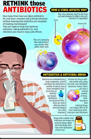
The next time you are down with viral fever, don’t rely on antibiotics to get better. You might have come back with a prescription for antibiotics after the doctor diagnosed you with a viral infection but rest assured they will not cure you.

rethink antibiotics Antibiotics are made specifically for the treatment of bacterial infections. Not only does taking antibiotics for a viral infection fail to cure it, the drug also ends up killing the harmless and protective bacteria in the body.

Says Dr C M Gulathi, editor, Monthly Index of Medical Specialities: ‘‘Prescribing antibiotics for viral is wrong as it serves no purpose. In fact, it can lead to harmful side effects. Most kinds of fever and throat infections are results of viral infection.’’

Most doctors are under immense pressure from the patient to prescribe drugs as medicines usually give a sense of security to the patient, making them feel better instantly. Explaining why doctors prescribe antibiotics for viral infections, Gulathi adds: ‘‘Doctors know that patients are going to start distrusting them if they don’t prescribe medicines. Secondly, doctors don’t spend too much time making the diagnosis these days. Prescribing antibiotics for viral infection is an outcome of poor diagnosis. Thirdly, doctors are also under pressure from private pharmaceutical companies to boost their business by prescribing their drugs.’’

   A virus is a small infectious agent that attaches itself to a living cell known as the host cell and starts reproducing. This leads to what we know as a viral infection. The virus attaches itself to the host cell and releases its DNA in the cell, forcing it to reproduce. The cell eventually dies as the virus prevents it from performing its normal functions. When it dies the cell releases new viruses that infect other cells. The most common viral infections are those of the nose, throat and the upper airways — in other words, upper respiratory infections. Sore throat, sinusitis, common cold and influenza are all caused by viruses.

   Viral infections are usually diagnosed through the symptoms presented. Accurate diagnosis can become difficult sometimes, in which case blood tests and cultures might be needed. Drugs that combat viral infections are known as antiviral drugs. Most antiviral drugs interfere with replication of the virus. These drugs include interferons, immunoglobulins and vaccines. Interferons stop viral infections. Immunoglobulin have antibodies which fight infection.

   There are ten times as many bacteria as human cells in our body. Most of these bacteria are harmless or protective to the immune system. Along with killing the pathogenic bacteria, which can cause infections, antibiotics can kill the bacteria needed by the body as well. It can also make the harmless bacteria immune to antibiotics.

   Antibiotics are not effective against viral infections but if a person has a bacterial infection in addition to a viral infection, an antibiotic is often necessary.

Source:TOI

Friday, June 13, 2008

New Way To Target And Kill Antibiotic-resistant Bacteria

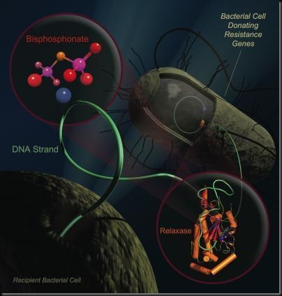
Putting bacteria on birth control could stop the spread of drug-resistant microbes, and researchers at the University of North Carolina at Chapel Hill have found a way to do just that. The team discovered a key weakness in the enzyme that helps "fertile" bacteria swap genes for drug resistance. Drugs called bisphosphonates, widely prescribed for bone loss, block this enzyme and prevent bacteria from spreading antibiotic resistance genes, the research shows. Interfering with the enzyme has the added effect of annihilating antibiotic-resistant bacteria in laboratory cultures. Animal studies of the drugs are now underway.

"Our discoveries may lead to the ability to selectively kill antibiotic-resistant bacteria in patients, and to halt the spread of resistance in clinical settings," said Matt Redinbo, Ph.D., senior study author and professor of chemistry, biochemistry and biophysics at UNC-Chapel Hill.

The study provides a new weapon in the battle against antibiotic-resistant bacteria, which represent a serious public health problem. In the last decade, almost every type of bacteria has become more resistant to antibiotic treatment. These bugs cause deadly infections that are difficult to treat and expensive to cure.

Every time someone takes an antibiotic, the drug kills the weakest bacteria in the bloodstream. Any bug that has a protective mutation against the antibiotic survives. These drug-resistant microbes quickly accumulate useful mutations and share them with other bacteria through conjugation -- the microbe equivalent of mating.

Conjugation starts when two bacteria smoosh their membranes together. After each opens a hole in their membrane, one squirts a single strand of DNA to the other. Then the two go on their merry way, one with new genes for traits such as drug resistance. Many highly-drug resistant bacteria rely on an enzyme, called DNA relaxase, to obtain and pass on their resistance genes. A mutation that provides antibiotic resistance can sweep through a colony as quickly as the latest YouTube hit.

The researchers analyzed relaxase because it plays a crucial role in conjugation. The enzyme starts and stops the movement of DNA between bacteria. "Relaxase is the gatekeeper, and it is also the Achilles' heel of the resistance process," Redinbo said.
Antibiotic Resistance

Led by graduate student Scott Lujan, the team suspected they could block relaxase by searching for vulnerability in a three-dimensional picture of the relaxase protein. Lujan, a biochemistry graduate student in the School of Medicine, confirmed the hunch using x-ray crystallography, which creates nanoscale structural images of the enzyme.

The researchers predicted that the enzyme's weak link is the spot where it handles DNA. Relaxase must juggle two phosphate-rich DNA strands at the same time. The team suspected a chemical decoy -- a phosphate ion -- could plug this dual DNA binding site. Redinbo, who has a background in cancer and other disease-related research, realized that bisphosphonates were the right-size decoy.

There are several bisphosphonates on the market; two proved effective. The drugs, called clodronate and etidronate, steal the DNA binding site, preventing relaxase from handling DNA. This wreaks havoc inside E. coli bacteria that are preparing to transfer their genes, the researchers found. Exactly how bisphosphonates destroy each bacterium is still unknown, Redinbo said, but the drugs are potent, wiping out any E. coli carrying relaxase. "That it killed bacteria was a surprise," he said. By targeting these bacteria, the drugs act like birth control and prevent antibiotic resistance from spreading.

Redinbo, who cautions that the results only apply to E. coli, said further testing will reveal whether bisphosphonates also attack similar species like Acinetobacter baumannii (hospital-acquired pneumonia), Staphylococcus aureus (staph infections) and Burkholderia (lung infections).

"We hope this discovery will help existing antibiotics or offer a new treatment for antibiotic-resistant bacteria," he said.

The drugs may be most effective at sites where clinicians can best control dosage on skin and in the gastrointestinal tract, Redinbo said. Other applications may include disinfectants and treatments for farm animals.

Source:Science Daily

Tuesday, June 10, 2008

Fungus That Produces Biofuels From Plants

The fungus Trichoderma reesei optimally breaks down plants into simple sugars, the basic components of ethanol. The fungus's genome has recently been sequenced by researchers from the Architecture et fonction des macromolécules biologiques laboratory (CNRS/Université de la Méditerranée and Universite de Provence), working together with an American team. The results show that only a few genes are responsible for the fungus's enzymatic activity. They offer new avenues for the fabrication of second generation biofuels from plant waste.

Fungus producing biofuels

The fungus Trichoderma reesei was discovered in the South Pacific during the Second World War, where it was damaging American military equipment and was defeating every attempt at protecting the equipment with cotton cloth. The fungus contains a number of enzymes, cellulases, with potent catalytic properties that break down plants.  It is considered to be the world’s most efficient fungus at breaking down the cellulose in plant cell walls into simple sugars, which it feeds on.

After fermentation, simple sugars can easily be transformed into biofuels such as ethanol. First generation agrofuels, made from grain or from beet, have certain limitations. Second generation biofuels, made from foresting and agricultural waste (tree cuttings, corn cobs, straw, etc.) do not have these limitations, as they complement pre-established agricultural activity, have a better CO2  balance, et don’t interfere with the agro-alimentary cycle.  To produce these second generation biofuels, industrialists are looking to develop fungus strains capable of producing a cocktail of cellulases and hemicellulases at a concentration of 50 g/l. Trichoderma reesei is the choice organism for most projects in this field.

Bernard Henrissat’s glycogenomic team at the Architecture et fonction des macromolécules biologiques lab specializes in the study of enzymes which break down sugars.* In order to learn more about the incredible enzymatic activity of Trichoderma reesei, they assayed its genome.  They found that the fungus has an unexpectedly small number of genes encoding cellulases (hemicellulases and pectinases), many fewer in fact than in usually found in fungi capable of breaking down plant cell walls. Moreover, the fungus has no or very little enzymatic activity allowing the digestion of specific components in the cell wall.

This was first interpreted as bad news, but the limitations of this model organism are now being seen as something positive. The fungus’s enzyme cocktail lends itself to numerous genetic modifications, and researchers are looking into which other enzymes can be added to the fungus’s gene sequence in order to make it even more efficient at producing bioethanol.

*The laboratory has a perfected Carbohydrate-Active Enzymes (CAZy) database which describes a number of enzyme families which form and destroy bonds between sugars.

Source:Science  Daily

Monday, June 9, 2008

Probiotics

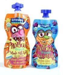
The original observation of the positive role of these bacteria can be credited to the pioneering work of Metchnikoff in the early 1900s, who suggested that these beneficial bacteria could be administered with a view to replacing harmful microbes with useful ones. The term "probiotic" meaning 'for life' was first coined in the 1960s by Lilly and Stillwell. Probiotics were defined as microorganisms proven to exert health-promoting influences in humans and animals. Probiotics were recently redefined by an expert group to be 'live microorganisms which when administered in adequate amounts confer a health benefit on the host'.

Probiotics

Organisms used as Probiotics

Two main genera of Gram-positive bacteria, Lactobacillus and Bifidobacterium, are used extensively as probiotics. However, while other probiotics such as Escherichia, Enterococcus and Saccharomyces are also available in the market, their safety remains an area of concern.

Benefits of Probiotic

  • People use probiotics to prevent diarrhea caused by antibiotics. Antibiotics kill "good" (beneficial) bacteria along with the bacteria that cause illness. A decrease in beneficial bacteria may lead to diarrhea. Taking probiotic supplements (as capsules, powder, or liquid extract) may help replace the lost beneficial bacteria and thus help prevent diarrhea. A decrease in beneficial bacteria may also lead to development of other infections, such as vaginal yeast and urinary tract infections, and symptoms such as diarrhea from intestinal illnesses
  • Helps to reduce Inflammation of the ileal pouch (pouchitis) that may occur in people who have had surgery to remove the colon.
  • Help prevent infections in the digestive tract.
  • Help control immune response (inflammation), as in inflammatory bowel disease (IBD), colon cancer, and irritable bowel syndrome (IBS).

Are probiotics safe?

Probiotic bacteria are already part of the normal digestive system and are considered safe. The U.S. Food and Drug Administration (FDA) does not regulate dietary supplements in the same way it regulates medication. A dietary supplement can be sold with limited or no research on how well it works or on its safety.

Conclusion

Literature on the role of probiotics in the treatment of pouchitis is still regarded as limited although small controlled trials have suggested that at least one probiotic preparation (VSL#3) containing 5 x 10 per gram of four strains of Lactobacilli , three strains of Bifidobacteria and one strain of Streptococcus salivarius subspecies thermophilus may be effective in the prevention of pouchitis.

Source:Google

Friday, June 6, 2008

Gold rush: Microbes with Midas touch

The tale of the hen that lay golden eggs is one almost all of us have heard. But Australian scientists have a new tale to tell — that of a ‘golden’ bacteria.

  Researchers in kangaroo country have uncovered evidence that a tiny microbeclip_image001 may have the Midas touch of Greek legend — capable of turning dust to gold. A bacteria known as Ralstonia metallidurans may play a key role in forming gold nuggets and grains. A group of scientists led by German-born researcher Frank Reith collected gold grains from two Australian mines more than 3,000 kilometres apart, and discovered that 80% of the grains had the bacteria living on them.

Gold Microbe

   “What we found out suggests that bacteria can accumulate this gold,” Reith told reporters during a telephone interview from his Australian office on Friday. Reith said Ralstonia metallidurans act as microscopic soil scrubbers, soaking up heavy metals in their dissolved form and converting them into less toxic, solid forms.

   “Heavy metals are toxic, not only to us but also to micro-organisms, in elevated concentrations,” he said. “It appears to be that the organism can detoxify its immediate environment of this toxic mobile gold and in this way gain a metabolic advantage. That’s why it would be present on these gold grains,” he elucidated.

   Many scientists have questioned the possible microbial role in forming gold, maintaining instead that gold grains were either remnants of larger pieces or formed through chemical processes over a period of time.

   Reith said his findings provide the strongest evidence yet that bacteria could play a key role in creating solid gold and could bring about a revolution in the entire industry in terms of its economics and revenue generation, although the exact mechanism is not yet known.

Source:TOI

Tuesday, June 3, 2008

We are 90% bacteria, actually

Humans Are Dependent On Microbe For Their Well-Being

We may not be entirely human, gene experts said after studying the DNA of hundreds of different kinds of bacteria in the human gut. Bacteria are so important to key functions such as digestion and the immune system that we may be truly symbiotic organisms – relying on one another for life itself, says Scientists. Their findings suggest that studying bacteria native to our bodies may provide important clues to disease, nutrition, obesity and how well drugs will work in individuals, said the team at The Institute for Genomic Research, commonly known as TIGR, in Maryland.

“We are somehow like an amalgam, a mix of bacteria and human cells. There are some estimates that say 90% of the cells in our body are actually bacteria,” Steven Gill, a molecular biologist formerly at TIGR and now at the State University of New York in Buffalo, said in a telephone interview. “We’re entirely dependent on this microbial population for our well-being. A shift within this population, often leading to the absence or presence of beneficial microbes, can trigger defects in metabolism and development of diseases such as inflammatory bowel disease.”

90% bacteria - Copy

Scientists have long known that at least 50% of human feces, and often more, is made up of bacteria from the gut. Bacteria start to colonise the intestines and colon shortly after birth, and adults carry up to 100 trillion microbesclip_image001[1], representing more than 1,000 different species.They are not just freeloading. They help humans to digest much of what we eat, including some vitamins, sugars, and fiber. They also synthesize vitamins that people cannot.

“Humans have evolved for million of years with these bacteria. And they provide essential functions,” Gill said. Gill and his team sequenced the DNA in feces donated by three adults. They found a surprising amount of it came from bacteria. They compared the gene sequences to those from known bacteria and to the human genome and found this socalled colon microbiome—the entire sum of genetic material from microbes in the lower gut—includes more than 60,000 genes. That is twice as many as found in the human genome.

  “Of all the DNA sequences in that material, only 1 to 5% of it was not bacterial,” Gill said. “We were surprised.” They also found a surprising number of Archaea, also known as archaebacteria, which are genetically distinct from bacteria but which are also one-celled organisms often found in extreme environments such as hot springs.   The donors were healthy adults. None had taken antibiotics for a year, as these drugs are known to disturb the bacteria in the body.

   Gill said his team hopes now to make a comparison of the gut bacteria from different people.

   “The ideal study would be to compare 20 people, 30 people from different ethnic backgrounds, different diets, drinkers, smokers, and so on, because I think there are going to be distinct differences,” Gill said.

   These bacteria almost certainly help break down drugs that people take and studying the effects of different populations of the microbes might provide clues to treating different people with various medications.

Source: TOI

Monday, June 2, 2008

Bacteria Mix in Guts of Babies Predicts Obesity

 The mix of bacteria in a baby's gut may predict whether that infant will become overweight or obese later in life, a new study suggests. Babies with high numbers of Bifidobacteria and low numbers of Staphylococcus aureus may be protected from excess weight gain, according to a team of researchers from the University of Turku in Finland.

 Bifidobacteria The help explain why breast-fed babies are at lower risk for later obesity, since Bifidobacteria are prevalent in the guts of breast-fed babies. Other studies repeatedly have found that being breast-fed is associated with a reduced risk of excess weight or obesity in childhood, with the risk lowered from 13 percent to 22 percent. In the new study, researchers evaluated children who had been part of a long-term study to evaluate the effect of probiotics on allergic disease. Probiotics are potentially beneficial bacteria found in foods such as yogurt and in dietary supplements.

The children had been evaluated at birth, five more times before age 2, and then again at ages 4 and 7. The researchers in the original study had also tested for intestinal microbes in fecal samples collected at 6 months and 12 months. For this latest study, the Finnish researchers selected 49 participants from the larger study -- 25 of them were overweight or obese at age 7 years, and 24 were normal weight at the same age. When they looked at the fecal samples, the average bacterial counts of Bifidobacteria when taken at 6 months and 12 months were twice as high in those who were a healthy weight as in those who got heavy. Those who stayed at a healthy weight also had lower fecal S.aureus levels at 6 months and 12 months than did those who got heavy.

Staphylococcus aureus

The S.aureus may trigger low-grade inflammation, the authors speculated, and that may also contribute to developing obesity. In other research, gut bacteria in adults have been found to be altered in obese adults who lost weight. Someday, the Finnish researchers speculated, tinkering with gut flora may help prevent or treat obesity.

The latest study doesn't pinpoint exactly why intestinal bacteria are linked with the development of obesity, said Connie Diekman, director of university nutrition at Washington University in St. Louis and president of the American Dietetic Association.

"The exact role that bacteria in the intestine play in development of obesity is still the subject of much research," she said, "but the benefits of breast-feeding are clear. Breast-feeding provides not only the proper nutrition for your infant, but it provides benefits that may impact long-term health and weight issues as well."

However, she added that, "while breast-feeding may play a role in the weight of children, so many other factors influence weight that parents shouldn't ignore good role modeling of healthy food choices, proper portions and regular physical activity. Healthy weight is a combination of factors, and no single issue will be the cause of weight gain or the magic answer to weight loss."

Another expert who has studied how obesity changes microbes in the gut calls the new study unique, because it collected information over several years and could look for differences in gut microflora. "The finding, that the lean children harbored higher levels of Bifidobacteria at younger ages, is very intriguing," says Ruth Ley, a research assistant professor at Washington University School of Medicine in St. Louis. Still, she says, research on the role of gut bacteria in regulating body weight is in the very early stages.
Source:Health Day News

__________________________________________________________________________