Wednesday, July 30, 2008

ATP Bioluminescence Techniques

 I began my career with project which was funded by Centre of Military Airworthiness & Defence Research & Development Organization, Bangalore.

Project was divided into two phases

1. Development of Bio kits for detection of Aviation Fuels.

2. Detection of Contamination in Aviation Fuels by Bioluminescence Techinque.

Phase I project we designed kits to detect contamination by methods like Peptone Dextrose Bromothymol blue, Dipslide technique, Bio Det test and Membrane Flitration Technique. I would brief about one test which we carried out in lab.

TTC

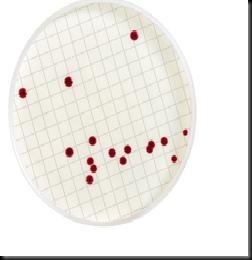
Bio Det Test: Redox dyes are being used to detect contamination. TTC (triphenyl tetrazolium chloride) is used. In the presence of metabolizing microbial cells TTC is converted to Formazone. This conversion in the medium is noted by change from characteristic colour of the medium to reddish pink. Change in colour is directly indicates presence of microbes and therefore fuel is not fit for consumption in aviations.

Phase II is Rapid Tests

Bioluminescence is the ability of living things to emit light. It is found in many marine animals, both invertebrate (e.g., some cnidarians, crustaceans, squid) and vertebrate (some fishes); some terrestrial animals (e.g., fireflies, some centipedes); some fungi and bacteria. The molecular details vary from organism to organism, but each involves a luciferin, a light-emitting substrate a luciferase, an enzyme that catalyzes the reaction ATP, the source of energy molecular oxygen, O2.

The more ATP available, the brighter the light. In fact, firefly luciferin and luciferase are commercially available for measuring the amount of ATP in biological materials.

The efficient light-producing enzyme system of ATP bioluminescence for the rapid detection of microorganisms. All actively respiring organisms contain adenosine triphosphate (ATP), which is used as the universal currency of free energy in biological systems. The luciferase enzyme hydrolyses ATP to AMP, releasing the stored energy as visible light. Luciferase can therefore be used to quickly detect the presence or absence of viable microorganisms in a sample by examining it for their ATP.

The mechanism of the light-producing reaction is:eq This light produced is measured using a instrument called "Luminometer". The intensity of light produced is proportional to the concentration of ATP and thus number of microbial cells.

Luminometer

The reaction is very efficient; every molecule of ATP can cause emission of a photon of green light. This provides a more rapid microbial detection system than waiting for visible colonies to grow on agar plates.

Some raw materials and end products may contain high levels of non-microbial ATP that can interfere with the bioluminescence reaction. This non-microbial ATP needs to be removed by use of an apyrase pretreatment.  ATP-depleting reagent  that is capable of isolating and removing both free and somatic ATP, enabling the detection of microbial ATP in contaminated biopharmaceutical samples.
Tuesday, July 29, 2008

Can Your Home Trigger Asthma?

Environmental Toxicologists Link Household Bacteria to Asthma

Scientists have found that chemicals called endotoxins can inflame airways and trigger asthma. Endotoxins are shed by bacteria in household dust. Experts say better home hygiene, washing bed linens in hot water at least once a week, and using allergen-prevention pillow cases and mattress covers can reduce the risk of asthma attacks.

Researchers say asthma and allergy triggers may be commonly found at home. That means there are things you can do to reduce the cause of your family's symptoms.

A new study indicates bacteria in household dust releases chemicals called endotoxins, which can inflame airways and trigger asthma.

endotoxin

Peter Thorne, an environmental toxicologist at the University of Iowa in Iowa City says, "If you think of a bacterium as an orange, the endotoxin is the material that makes up the peel. It's the outer layer. And this becomes shed from bacteria, and it's everywhere in the environment.

Researchers found the bedroom had fewer endotoxins, but it had a greater impact on a child's health. After all, that's where kids spend almost half their day. Plus, they have closer contact with the endotoxins. "Endotoxin in the home is related to higher rates of asthma," Thorne says.

There are things you and your family can do to lower the amount of endotoxins: wash your bed linens in hot water at least once a week, reduce clutter so you can clean better, purchase allergen-prevention pillow cases and mattress covers and don't eat in bed.

"I just try and be a little more conscious inside of what I can do so I can control my environment on the inside," Pannkuk tells DBIS. He is stuck with his seasonal allergies but is happy he can control environmental factors that affect his family.

Another cause of asthma in the home is pet dander. Cases of asthma tripled among newborns to 4-year-olds between 1980 and 2000.

BACKGROUND: Bacteria lurking in household dust produces chemicals that may trigger asthma symptoms, whether a person suffers from asthma or allergies or not. The results, from the first nationwide study of toxins made by bacteria in households, indicate that it's not just the concentration of the bacteria-made toxin that is important. Other factors, such as how long and when a person is exposed to the bacterial toxin, as well as genetic factors, may contribute to the development of asthma.

THE STUDY: The nationwide study involved the analysis of more than 2,500 dust samples from 831 homes across the U.S. Results showed a strong association between the levels of toxins made by bacteria -- called endotoxins -- and the prevalence of diagnosed asthma, asthma symptoms and wheezing. People in households with higher endotoxin concentrations had higher instances of respiratory symptoms.

ABOUT ENDOTOXINS: Endotoxins are found in the cell wall of bacteria and are only released when the cell ruptures or disintegrates. Because bacteria can be found everywhere in the home, the likelihood of such a release is very high. Once released, endotoxins can cause inflammation of the airways and lead to asthma symptoms. Endotoxin levels can be reduced in the same way that allergy symptoms can be reduced: by removing dust, keeping the floors as clean as possible, keeping moisture low, repairing water damage, cleaning bed linens, and using a high-efficiency vacuum cleaner with a HEPA filter, among other actions.

ASTHMA OR ALLERGIES? Asthma is a chronic disease affecting the airways that carry air in and out of the lungs. The inside walls of the airways become inflamed (swollen) and narrower so less air can flow through the lung tissues. This in turn causes wheezing, coughing, tightness in the chest, and trouble breathing. Asthma is linked to allergies, although not everyone with asthma has allergies. People with allergies tend to react more strongly to the presence of allergens such as animal dander, dust mites, pollen or mold, as well as cigarette smoke and air pollution.

Source: ScienceDaily

Monday, July 28, 2008

Watch your step: E. coli can lurk at the beach

Potentially dangerous bacteria found near freshwater spots nationwide

The perils of a day at the beach aren’t always as easy to see as riptides, broken shells and jellyfish — the sand at the shore may harbor E. coli and other potentially dangerous disease-causing bacteria, a recent study showed.

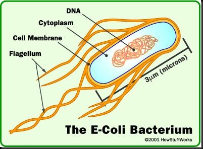
E. coli is one of the main species of bacteria that live in the lower intestines of mammals, including humans — one person excretes billions of them in a day. Pathogenic strains of E. coli can cause vomiting and diarrhea.

E.coli

Government testers look for E. coli as an indicator of fecal contamination at freshwater beaches all over the country, because the other microbes present are more difficult to detect (another bacteria is used to test for fecal matter at ocean beaches because E. coli does not survive well in salt water).

Beaches all over the country frequently close due to fecal contamination; a day at the beach can be ruined if septic systems overflow or malfunction, or if a lot of birds happen to be in the neighborhood.

Seasonal sources
To test exactly which
strains of E. coli were sitting in the sands around Lake Superior, and whether any of them were potentially dangerous to humans, a group of University of Minnesota researchers collected samples and compared the DNA to an existing library.

Their results are detailed in a recent issue of the journal Environmental Science and Technology.

They found two broad types of E. coli in the sand: those “deposited more recently,” as team member Michael Sadowsky put it, and those “that have learned to kind of grow or reproduce in the sand,” he said.

The levels of both of these sources vary seasonally. Those that have become indigenous to the lake sands tend to be more abundant in the summer, when nutrients are more available and temperatures rise. Contributions from birds tend to come when they are migrating through the area. Fecal contamination from sewage can occur whenever there is a malfunction or overflow.

More important, the study found that very few of the E. coli present on the beach are potentially harmful to humans — other microbes that tend to travel in the same waste streams, such as salmonella, you likely wouldn’t get sick just from wiggling your toes in the sand, because most of these bacteria follow what Sadowsky terms the “fecal to oral route.” Which means you should listen to your parents and wash your hands frequently, especially after using the bathroom and before eating.

“Getting it on your skin is not going to be very dangerous,” he said.

Just watch what you swallow.

Source:Msnbc

Friday, July 25, 2008

Eye Ointment Can Cure Frog Fungus

An antibiotic primarily used to treat pinkeye in humans rids frogs of the fungal disease that is wiping out amphibian populations around the world, a team of New Zealand scientists reports.

Infected frogs treated with the drug for two weeks were cured of the deadly disease, called chytridiomycosis or frog chytrid disease.

frog fungus

The fungal disease, along with habitat destruction and global warming, is blamed for putting more than a third of the world's 6,234 known amphibian species on the path to extinction.Scientists have been racing to find a way to curb the disease before it takes an even heavier toll. The antibiotic chloramphenicol is "definitely the best" treatment found to date, Bishop said.

Another drug, itraconazole—which is used to treat fungal and yeast infections in humans—has also been shown to cure chytrid disease. But the drug causes kidney damage in some frogs, he noted.

Itraconazole also remains under patent protection and is therefore expensive, whereas chloramphenicol is generic and widely available.

Captive Tests

Allan Pessier is a veterinary pathologist at the San Diego Zoological Society in California and an expert on the deadly fungal disease. He said more tests are needed before declaring one drug better.

Both drugs, he added, are effective treatments for captive frogs but are probably never going to see widespread use in the wild as a spray or liquid dumped into streams and ponds.

"It's a step that helps us manage the disease, but it's not like declines associated with chytridiomycosis are going to go away because of this discovery," he said.

To be effective in the wild, scientists would need to overcome hurdles like maintaining sufficient concentrations of the drug in the environment without harming the ecosystem, he noted.

Bishop's colleague, biochemist Russell Poulter of Otago, stumbled upon chloramphenicol while running a battery of tests on various antibiotic and antifungal compounds in search of any that may prove effective against chytrid disease.

"It was a very surprising result to find an antibacterial that killed a fungus," Bishop said.

The researchers used the compound both as an over-the-counter ointment applied to infected frogs' skin and as a solution soaked into a paper towel on which the frogs sat.

"We found that after about 18 days we could swab the frogs again and they would be cured of chytrids," Bishop said.

The team tested the compound on three species in New Zealand: the brown tree frog and the southern bell frog—which were introduced from Australia—and the critically endangered native Archey's frog.

Bishop said all the frogs are still alive and well 12 months after treatment. Researchers at the Taronga Zoo in Sydney, Australia, are now testing the drug on six more endangered frog species.

Why the drug kills the fungus, he added, remains under investigation.

Drug Resistance?

The New Zealand researchers also tried to re-infect frogs that had been successfully treated. The frogs again contracted the disease, but it disappeared within a few days.

Bishop is uncertain why the frogs were "difficult to re-infect." Perhaps the chloramphenicol lingers in their skin or it works like a vaccine, priming the amphibian immune system to thwart the fungus, he said.

"We're hoping this is going to be something really significant, and we are currently investigating that," he added.

Reid Harris is a biologist at James Madison University in Harrisonburg, Virginia, who studies chytrid disease. He agreed further studies are needed to investigate the apparent disease resistance.

"The more we can learn about any sort of mechanism of resistance ... the better," he said.

Conserving Frogs

Bishop and his colleagues are continuing to comb through various antifungal agents in search of one that is effective against chytrid disease.

Antifungals, he noted, may prove more acceptable to spraying in the wild than chloramphenicol or itraconazole.

"People don't want antibiotics being dumped in vast quantities in the environment," he said.

Meanwhile, Pessier of the Zoological Society of San Diego said the antibiotics can be used to treat and maintain captive populations until a solution to the problem in the wild is found.

"If you didn't have that option," he said, "you'd see frogs going essentially extinct because of the infection."

Source:National Geographic

Wednesday, July 23, 2008

Overweight? Blame the bacteria in your gut

'Microbial component' may contribute to obesity, research indicates

The size of your gut may be partly shaped by which microbes call it home, according to new research linking obesity to types of digestive bacteria.

Both obese mice — and people — had more of one type of bacteria and less of another kind, according to two studies published Thursday in the journal Nature.

A “microbial component” appears to contribute to obesity, said study lead author Jeffrey Gordon, director of Washington University’s Center for Genome Sciences.

Obese humans and mice had a lower percentage of a family of bacteria called Bacteroidetes and more of a type of bacteria called Firmicutes, Gordon and his colleagues found.

bacteria gut

The researchers aren’t sure if more Firmicutes makes you fat or if people who are obese grow more of that type of bacteria.

But growing evidence of this link gives scientists a potentially new and still distant way of fighting obesity: Change the bacteria in the intestines and stomach. It also may lead to a way of fighting malnutrition in the developing world

Nikhil Dhurandhar, a professor of infection and obesity at Louisiana State University’s Pennington Biomedical Research Center, wasn’t part of the research, but said it may change the way obesity is treated eventually.

“We are getting more and more evidence to show that obesity isn’t what we thought it used to be,” Dhurandhar said. “It isn’t just (that) you’re eating too much and you’re lazy.”

He said the field of “infectobesity” looks at obesity with multiple causes, including viruses and microbes. In another decade or so, the different causes of obesity could have different treatments. The current regimen of diet and exercise “is like treating all fevers with one aspirin,” Dhurandhar said.

In one study, Gordon and colleagues looked at what happened in mice with changes in bacteria level. When lean mice with no germs in their guts had larger ratios of Firmicutes transplanted, they got “twice as fat” and took in more calories from the same amount of food than mice with the more normal bacteria ratio, said Washington University microbiology instructor Ruth Ley, a study co-author.

It was as if one group got far more calories from the same bowl of Cheerios than the other, Gordon said.

In a study of a dozen dieting people, the results also were dramatic.

Before dieting, about 3 percent of the gut bacteria in the obese participants was Bacteroidetes. But after dieting, the now normal-sized people had much higher levels of Bacteroidetes — close to 15 percent, Gordon said.

“I think that gut bacteria affects body weight,” said Virginia Commonwealth University pathology professor Richard Atkinson, who wasn’t part of the research team and is president of Obetech Obesity Research Center in Richmond. “I don’t think there’s any doubt about that and they showed that.”

Most microbes beneficial
The growing field of research puts more importance in the trillions of microbes that live in our guts and elsewhere, crediting it with everything from generations of people getting taller to increases in diabetes and asthma.

People are born germ-free, but within days they have a gut blooming with microbes. The microbes come from first foods — either breast milk or formula — the exterior environment, and the way the babies are born, said Stanford University medicine and microbiology professor David Relman, who was not part of the study.

For decades, doctors have treated bacteria in a “warlike” manner, yet recent research shows that “most encounters we have with microbes are very beneficial,” Gordon said.

“Much of who we are and what we can do and can’t do as human beings is directly related to microbial inhabitants,” Relman said.

Source:msnb

Monday, July 21, 2008

Resistant Microbes Behind Soft Contact Lens’ Corneal Infections

A new study from University Hospitals Case Medical Center has revealed the mechanism through which fungus strains cause corneal infections, similar to the ones that compelled ‘Bausch & Lomb’ to withdraw its ReNu with MoistureLoc contact lens solution in 2006.

The researchers have shown that these infections were fuelled and made resistant to treatment by the formation of a highly resistant structure of microbial cells held together with a glue-like matrix material. They call this conglomeration of cells biofilms.

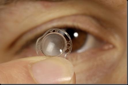
lenses
“Once they live in that type of state, the cells become resistant to lens solutions and immune to the body’s own defence system. This study should alert
contact lens wearers to the importance of proper care for contact lenses to protect against potentially virulent eye infections,” said senior researcher Mahmoud A. Ghannoum, director of the Center for Medical Mycology at University Hospitals Case Medical Center.

“Biofilms are a constellation of resistant organisms,” he added. This is the first study to provide in-vitro model for cornea keratitis infections caused by a fungus called Candida albicans, and another fungus Fusarium, which was one of the main culprits in the ReNu with Moisture Loc cases.

Dr. Ghannoum says that the study has also revealed that the strain of fungus (ATCC 36031) used for testing the effectiveness of lens care solutions is a strain that does not produce biofilms as the clinical fungal strains do. Lens care solutions, which are currently tested against an old and rare genotype fungal strain obtained in the 1970s from a patient from Nigeria, are effective in the laboratory, but fail when faced with strains in real-world situations.

“The multipurpose contact lens solutions cannot kill these germs, to put it simply,” said Dr. Ghannoum.

“We recommend that solutions be tested for biofilms produced by more recent clinical isolates. One of the underlying reasons for ReNu with MoistureLoc not being effective against the outbreak of keratitis is that the solution is not effective against biofilms and the organisms contained in biofilms,” he added.

During the study, six kinds of contact lenses made by three different manufacturers and two lens care solutions—Bausch & Lomb’s MoistureLoc and MultiPlus—were tested against three fungal strains of Fusarium and one strain of C. albicans. Given the commoness of biofilm contamination of contact lens cases, and since ReNu with MoistureLoc is ineffective against fungal biofilm, Dr. Ghannoum says that the industry must ensure that their multipurpose solutions are effective against biofilms.

He also recommends exercising extreme caution to ensure the contact lens care case is not contaminated with biofilm. Dr. Ghannoum suggests that soft contact lens wearers use only fresh lens care solution, change the solution regularly, and avoid using the solution beyond the expiry date written on the package. He also recommends a rub and rinse method to clean contact lens, besides ensuring that the bottle caps and tips of multipurpose solutions are clean.

Source: Medindia.

Friday, July 18, 2008

Vitamin D3 Protects Skin from Harmful Microbes

A study shows that fluctuations in Vitamin D3 levels control the body’s innate immune response, affecting a skin wound’s ability to heal.

Richard L. Gallo, M.D., Ph.D., professor of medicine and chief of UCSD’s Division of Dermatology and the Dermatology section of the Veterans Affairs San Diego Healthcare System, says that several unexpected associations between fluctuations of the body’s vitamin D3 and infectious disease have emerged in recent investigations.

In a study appearing online February 8 in advance of publication in the March issue of the Journal of Clinical Investigation, Gallo and his colleagues look at how the innate immune system is controlled in the skin, and find that genes controlled by active vitamin D3 play an essential role in the process.
“Our study shows that skin wounds need vitamin D3 to protect against infection and begin the normal repair process,” said Gallo. “A deficiency in active D3 may compromise the body’s innate immune system which works to resist infection, making a patient more vulnerable to microbes.”

vitamin d

Gallo’s lab discovered that an antimicrobial peptide called cathelicidin is produced by wounds and is necessary to fight infections. Recently, several studies have begun to link vitamin D to cathelicidin. Researchers focused on white blood cells called macrophages that work to destroy invading bacterial microbes. Macrophages contain toll-like receptors that identify the invaders; when the receptors sense the presence of bacteria, they trigger cathelicidin production.

Gallo’s team has now discovered that injury stimulates skin cells called keratinocytes, which surround the wound, to increase the production of vitamin D3 and that this in turn increases the expression of genes (CD14 and TLR2) that detect microbes. These genes, together with active vitamin D3, called 1,25D3, then lead to more cathelicidin. In both mice and humans, a deficiency in cathelicidin allows infections to develop more readily.

“Our finding – that multiple, diverse genes controlled by 1,25D3 are increased after injury to the skin – suggests that the availability of D3 is essential to the wound. These responses are a previously unrecognized part of the human injury response,” said Gallo.

Lower concentrations of 1,25D3 in African Americans, likely due to a decreased ability to absorb vitamin D from sunlight, correlate with increased susceptibility to infection. In addition, 1,25D3 has been suggested to be an immune-modifying agent in pulmonary tuberculosis.

As a result of this and previous studies, Gallo and his colleagues are beginning clinical trials at UCSD Medical Center with both oral and topical vitamin D3. Normal volunteers, and patients with disorders in antimicrobial peptide production such as atopic dermatitis and acne, are being studied to determine if vitamin D3 can improve their natural immune defenses.

Source:Medindia

Thursday, July 17, 2008

Honey eliminates stubborn skin microbes

Medical grade honey may be used to prevent or treat infections of skin, burns, catheters and other skin-penetrating medical devices.

Medical grade honey kills antibiotic-resistant bacteria grown in the test tube and eradicates antibiotic-resistant bacteria "colonies" on the skin of healthy volunteers, Dutch researchers report.

honey 2

Given these findings, senior investigator Dr Sebastian A.J. Zaat said, "Medical grade honey might be used to prevent or treat infections of skin, burns, catheters and other skin-penetrating medical devices."

Honey, they point out, has been used since ancient times to successfully treat infected wounds. Moreover, there have been no reports of antibiotic resistance to honey.

However, large variation in the antibacterial property of various honeys has hampered current medical acceptance.

To investigate further, the researchers tested the antibacterial activity of the medical grade honey called Revamil (Bfactory), which is produced in greenhouses under standardised conditions.

They found that antibiotic-susceptible and antibiotic-resistant isolates of several common bacteria, including Staphylococcus aureus and E. coli, were killed within 24 hours after incubation with the honey.

They also found that the honey decreases the ability of these microbes to "colonise" the skin of healthy volunteers.

After applying honey for 48 hours to "bacteria-laden" patches of forearm skin, the extent of skin colonisation was reduced 100-fold.

Moreover, 81 per cent of the honey-treated skin patches yielded negative skin culture results compared to 21 per cent of control patches.

Following these encouraging results, continued Zaat, "We are presently investigating whether honey can be used to prevent line sepsis in intensive care patients. This potentially life - threatening infection is often caused by bacteria from the skin, which can be eradicated with honey."

Source:Mindfood

Monday, July 14, 2008

Red Wine can Protect People from Microbes

Wine lovers have one more reason to cheer. A US study reveals that drinking red wine and red grape juice may protect people from common food-borne illnesses.

The findings were recently presented at the Institute of Food Technologists annual conference in Chicago. Researchers at the University of Missouri-Columbia conducted studies in which they examined the effects of reseveratrol — the compound responsible for wine's red colouring — as well as ethanol and pH levels on common food pathogens such as E. coli, Salmonella typhimurium, Listeria monocytogenes and H. pylori.

They discovered that in addition to ethanol, reseveratrol and pH may inhibit the pathogens.  In the study, Cabernet, Zinfandel and Merlot wines were found to be particularly effective in defending against food-borne pathogens and had high anti-microbial properties.

Red wine

Yet the wines didn't have a negative effect on probiotic bacteria, which fight harmful bacteria in the intestines."This study showed that the four probiotics tested weren't inhibited by red wines; the pathogens were," said Azlin Mustapha, associate professor of food science at the University of Missouri-Columbia.

Numerous white wines also were tested, but yielded no positive results, the researchers said. "It's not just ethanol in the red wine that is inhibitory toward food-borne pathogens, but other factors which include the pH of the wine — because wines are a little acidic, and possibly the phytochemicals may have an effect," said Mustapha, noting that grape juice produces similar results.

"We hypothesize that these phytochemicals, reseveratrol being the main one, also play a role not just as antioxidants but also may have some inhibitions against food-borne pathogens."

Source: medindia.com

Friday, July 11, 2008

Rust-Breathing Bacteria: Miracle Microbes

They breathe rust, clean up polluted groundwater, generate electricity, and may harbor clues to the origins of life. That's a lot for one family of microscopic bugs, but don't be surprised when Derek Lovley wows the world with another wonder from the Geobacter genus of bacteria.

"When we think we have hit the last of the big discoveries, something else comes along," said Lovley, a microbiologist at the University of Massachusetts, Amherst.

The microbes' metabolism is unique: They use metals to get energy from food in the same way humans use oxygen. In 1987 Lovley discovered Geobacter metallreducens in some iron-rich mud he scooped up from the Potomac River.

Geobacter

In the 17 years since that discovery, Lovley and his colleagues have found more than 30 species in the bug family, sequenced the genomes of several, and filled scientific journals with the details of new insights.

Lovley collaborates with scientists around the world, and his lab buzzes with more than 50 researchers. Most recently, Lovley and his colleagues sequenced the genome of a Geobacter species that can generate electricity and cleanup groundwater contaminated with uranium.

"Never underestimate the abilities of the microbial world," said Tim Magnuson, a microbiologist who worked in Lovley's lab before moving to Idaho State University in Pocatello.

Magnuson believes that the Geobacter species and a number of other metal-reducing bacteria discovered since Lovley's seminal 1987 find are the just the beginning of what promises to be an exciting scientific journey into the world or microbes with unique metabolisms.

"The more we look, the more we discover. We have and will discover organisms that are not at all related to Geobacter but can carry out that kind of metabolism. It is a huge frontier."

Lovley Bugs

When Lovley embarked on his academic journey into the sciences, all he wanted to find was a good job in the outdoors. But as he pondered what made the world go around as an undergraduate biology major at the University of Connecticut in the 1970s, things began to change.

"It started to be apparent that these microorganisms account for a lot of biomass [living matter] on Earth and are big drivers for the environmental and ecological processes," Lovley said. "So I went to grad school and tried to learn microbiology."

After receiving his Ph.D. from Michigan State University in 1982, Lovley took a job with the United States Geological Survey studying how microorganisms affected groundwater in the Chesapeake Bay.

He knew that microorganisms could live in oxygen-deprived environments by processing things such as sulfate and methane. If they could do that, Lovely reasoned, they might also be able to make a living from iron. Which is why, in 1987, Lovely scooped up the iron-rich mud from the Potomac River just downstream from Washington, D.C. Unbeknownst to him, the microbes therein would change his life.

Back in his lab, Lovley put the mud in a test tube, added some acetate—essentially vinegar, a favorite microbe food—and watched. Eventually, he noticed little black minerals collecting on the bottom of the tube amidst a sea of fluffy orange iron oxide, or rust.

"I put a magnet up to the side of the tube, and all the little pieces flew over to the side of the glass," Lovely said. "That was a 'Eureka!' moment."

The mud-dwelling microbes, which Lovley later named Geobacter metallreducens, obtained their energy by transferring electrons onto the rust. In the process, they turned the rust into magnetite.

Magnetite is the source of most of the magnetic material deposited on Earth some two billion years ago. Lovley and his colleagues concluded in a 1987 paper published in Nature that the microbes may have been responsible for most of these early magnetite deposits.

Microbes in Action

Since Geobacter metallreducens was discovered, Lovley and his colleagues have tried to figure out how to take advantage of this unique microbial metabolism, from cleaning up contaminated groundwater to generating electricity.

Magnuson, the Idaho State University microbiologist, believes that the ability of microorganisms like Geobacter to clean up groundwater may be of particular use to humans in the coming decades, as world populations are predicted to experience a water crisis.

He envisions using microbes not only to decontaminate water supplies but to protect them until human consumption.

"Using Geobacter to protect and maintain the quality of these aquifers might be a viable solution, seeing as we are going to run into water-quality issues," he said.

Lovley and his colleagues are not yet developing Magnuson's proposed superefficient water filters, but they are actively developing a method to remove uranium from billions of gallons of contaminated groundwater.

Source:National geographic

Thursday, July 10, 2008

Scientists discover way to protect against death following plague infection

Bacteria that cause pneumonic plague can evade our first-line defences, making it difficult for the body to fight infection. In fact, a signature of the plague is the lack of an inflammatory response.

Now, scientists have discovered a way to protect against death following infection with plague bacteria, by using molecules that can mimic the pathogens. According to research published in the July issue of Microbiology, these molecules make antibiotics more effective and can even be used to protect against other diseases.

The plague, caused by Yersinia pestis, has killed an estimated 200 million people worldwide. Although treatments have improved, it remains a threat to public health. It can be transmitted from human to human in aerosols and is therefore listed as a Category A bioterrorism agent.

Yersinia pestis

"Yersinia pestis is successful in causing disease in mammals because it can dampen the normal non-specific immune response to infection," said Dr Scott Minnich from the University of Idaho, USA. "We found an intranasal therapy that stimulates the innate immune response and protects against pneumonic plague."

Following infection, lipid A (which is part of the bacterial surface) binds to receptors on our immune cells, triggering an immune response. Yersinia pestis circumvents this, stopping our cells from taking action. Molecules have been developed that mimic lipid A, eliciting a strong immune response that can prevent death in infected animals. Dr Minnich and his colleagues studied the effect of a nasal spray containing two such molecules, CRX-524 and CRX-527, on mice infected with Yersinia pestis.

"Treatment with synthetic modified lipid A molecules can directly protect animals against pneumonic plague infections," said Dr Minnich. "We also found that stimulating innate immunity using this nasal spray enhanced conventional antibiotic therapy. When it is given along with antibiotics, fewer doses and less antibiotic protects against pneumonic plague."

The results of this study suggest that synthetic modified lipid A compounds may provide a new therapeutic tool against plague infection. In a control group that did not receive the treatment, only 23% of mice survived for 3 days. When given the mimic molecules, up to 93% of mice survived for 3 days, 70% for 4 days and 34% recovered completely. This highlights the importance of the non-specific, first-line immune defences during the critical early phase of infection. Stimulating this response can over-ride a microorganism's counter measures to evade or disable the immune response.

Other studies have shown related therapeutic compounds are also effective against influenza and Listeria monocytogenes. "This work is still at a very basic animal model testing stage with regards to plague," said Dr Minnich. "What is exciting is that these studies provide insight into bacterial/host interactions in the disease process and promise new strategies to combat a variety of infectious agents."

Source: news-medical.net.

Wednesday, July 9, 2008

What Genes Help Microbes Invade Leafy Greens?

When unwanted microbes form an attachment, the consequences—for us—can be serious.

That’s if the microbes happen to be human pathogens like Listeria monocytogenes, if the target of their attentions happens to be fresh vegetables often served raw, such as cabbage or the sprouted seeds of alfalfa.

image

Scientists don’t yet fully understand how the malevolent microbes form colonies that cling stubbornly to and spread across plant surfaces, such as the bumpy leaves of a cabbage or the ultra-fine root hairs of a tender alfalfa sprout.

But food safety researchers at the ARS Western Regional Research Center in Albany, California, are putting together pieces of the pathogen puzzle.

A 1981 food-poisoning incident in Canada, caused by L. monocytogenes in coleslaw, led microbiologist Lisa A. Gorski to study the microbe’s interactions with cabbage. Gorski, with the center’s Produce Safety and Microbiology Research Unit, used advanced techniques not widely available at the time of the cabbage contamination.

“Very little is known about interactions between Listeria and plants,” says Gorski, whose study revealed the genes that Listeria uses during a successful cabbage-patch invasion.

The result was the first-ever documentation of Listeria genes in action on cabbage leaves. Gorski, along with coinvestigator Jeffrey D. Palumbo—now with the center’s Plant Mycotoxin Research Unit—and others, documented the investigation in a 2005 article in Applied and Environmental Microbiology.

Listeria, Behaving Badly

“People had looked at genes that Listeria turns on, or ‘expresses,’ when it’s grown on agar gel in a laboratory,” says Gorski. “But no one had looked at genes that Listeria expresses when it grows on a vegetable.

“We were surprised to find that when invading cabbage, Listeria calls into play some of the same genes routinely used by microbes that are conventionally associated with plants. Listeria is usually thought of as a pathogen of humans. We hadn’t really expected to see it behaving like a traditional, benign inhabitant of a green plant.

“It’s still a relatively new face for Listeria, and requires a whole new way of thinking about it.”

In related work, Gorski is homing in on genetic differences that may explain the widely varying ability of eight different Listeria strains to successfully colonize root hairs of alfalfa sprouts—and to resist being washed off by water.

Source:USDA

Friday, July 4, 2008

New Window Opens On The Secret Life Of Microbes

Nowhere is the principle of "strength in numbers" more apparent than in the collective power of microbes: despite their simplicity, these one-cell organisms--which number about 5 million trillion trillion strong (no, that is not a typo) on Earth--affect virtually every ecological process, from the decay of organic material to the production of oxygen.

But even though microbes essentially rule the Earth, scientists have never before been able to conduct comprehensive studies of microbes and their interactions with one another in their natural habitats. Now, a new study provides the first inventories of microbial capabilities in nine very different types of ecosystems, ranging from coral reefs to deep mines.

new

"These new microbial inventories provide a new and important window into ecosystems and how they respond to stresses, such as pesticide runoff and invasive species," said Lita Proctor, an NSF program director.

Rather than identifying the kinds of microbes that live in each ecosystem, the study catalogued each ecosystem's microbial "know-how," captured in its DNA, for conducting metabolic processes, such as respiration, photosynthesis and cell division. These microbial catalogues are more distinctive than the identities of resident microbes. "Now microbes can be studied by what they can do not who they are," said Proctor.

This microbial study employed the principles of metagenomics, a powerful new method of analysis that characterizes the DNA content of entire communities of organisms rather than individual species. One of the main advantages of metagenomics is that it enables scientists to study microbes--most of which cannot be grown in the laboratory--in their natural habitats.

Specifically, the microbial study produced the following results:

A unique, identifying microbial fingerprint for each of nine different types of ecosystems. Each ecosystem's fingerprint was based on its unique suite of microbial capabilities.

Methods for early detection of ecological responses to environmental stresses. Such methods are based on the principle that "microbes grow faster and so respond to environmental stresses more quickly than do other types of organisms," said Forest Rohwer of San Diego State University, a member of the research team. Because microbes are an ecosystem's first-responders, by monitoring changes in an ecosystem's microbial capabilities, scientists can detect ecological responses to stresses earlier than would otherwise be possible--even before such responses might be visibly apparent in plants or animals, Rohwer said.

Evidence that viruses--which are known to be ten times more abundant than even microbes--serve as gene banks for ecosystems. This evidence includes observations that viruses in the nine ecosystems carried large loads of DNA without using such DNA themselves. Rohwer believes that the viruses probably transfer such excess DNA to bacteria during infections, and thereby pass on "new genetic tricks" to their microbial hosts. The study also indicates that by transporting the DNA to new locations, viruses may serve as important agents in the evolution of microbes.

Source: ScienceDaily

Tuesday, July 1, 2008

Fighting Cancer With Salmonella

Disease-causing bacteria can help in the fight against cancer. This may sound a little far-fetched at first, but in future bacteria could form the basis for innovative tumor therapies. Researchers at the Helmholtz Centre for Infection Research in Braunschweig have succeeded in planting "remote-controlled" salmonella in the tumors of cancer-bearing mice. The genetically modified microbes can produce substances on command. "Perhaps at some point," hopes Helmholtz scientist Dr. Holger Loessner, "we will be able to make these bacteria secrete cell toxins precisely where they are needed: in the middle of cancerous tissue."

fight cancer

Dr. Loessner's hopes are based on a curious phenomenon that researchers have known about since the mid-19th century. When tumor patients encounter a bacterial infection, frequently tumors are colonized by the bacteria and provide a niche for their multiplication. What exactly triggers this behavior is not well understood. "We suspect that the dead tissue inside the tumor provides these bacteria with a protective and nutrient-rich environment, and therefore, attracts them," explains Dr. Siegfried Weiss, head of the project group "Molecular Immunity" at the Helmholtz Centre for Infection Research. "Tumor interiors," he adds, "are low in oxygen, conditions under which many types of bacteria thrive."

For Weiss, Loessner and their colleagues, more important than the basic principles are the possibilities this phenomenon opens up. The rush by bacteria to colonize tumors suggests this behavior could be used for human benefit. Weiss and Loessner have now shown that this is essentially feasible. For the first time, they inserted in bacteria of the genus Salmonella typhimurium, a gene cluster which can be "switched on" by administration of a simple sugar molecule called L-arabinose.

Applying L-arabinose, after the salmonella bacteria have infected mice and migrated to the cancerous tissue, activates these genes. Thus far, Weiss and Loessner used genes that encode light emitting proteins. If the mice are given a dose of the sugar, bacteria that have colonized the tumor fluoresce, such that the location and size of the tumor can be analyzed. Theoretically, instead of emitting light, the bacteria in future could be triggered to produce and deliver cancer-fighting medication inside the tumor.

Alternatively, they could also deliver immune-stimulating substances that would mark the tumor for the body's own immune-defense mechanisms and induce a salutary immune response. "For medical purposes, of course," notes Dr. Loessner, "we would not use dangerous pathogens, but rather mutant strains that are harmless to humans."

"Until now, people have viewed salmonella as a threat to their health," says project leader Weiss, "so there would be a certain charm in using such bacteria for treatment of such a terrible disease as cancer."

Source: Sciencedaily