Thursday, October 23, 2008

Advance Offers Revolution In Food Safety Testing

Microbiologists at Oregon State University have developed a new technology to detect illness-causing bacteria – an advance that could revolutionize the food industry, improving the actual protection to consumers while avoiding the costly waste and massive recalls of products that are suspected of bacterial contamination but are perfectly safe.

The new approach - made possible by fundamental research on the color changes in pigment-bearing cells from Siamese fighting fish - should be easier to use, faster and more directly related to toxicity assessment than conventional approaches now used to test food for bacterial contamination and safety.

"Rapid methods are not readily available to directly assess the toxicity of bacterial contamination in a user-friendly fashion," said Janine Trempy, professor of microbiology and associate dean of the OSU College of Science. "When this new technology is commercially available, we should be able to provide a higher level of assurance to the consumer while avoiding the waste of millions of dollars worth of food that is suspected of bacterial contamination, but actually is safe."

Bacterial illnesses associated with food and water can produce symptoms ranging from mild stomach upset to severe illnesses and even death, and they are common. It's been estimated there are about 76 million illnesses of this type every year that cost the U.S. more than $10 billion.

Part of the problem is that conventional food safety testing done with DNA-based tests or antibody-based methods only indicate the presence of specific bacteria, which does not necessarily describe toxicity and the potential to cause harm. Sometimes bacteria only exhibit the behavior that can cause illness under specific environmental conditions, and it's that toxic behavior that we need to detect, Trempy said.

"Bacteria are common on exposed surfaces, including the food products we consume," Trempy said. "Simply knowing they are there doesn't completely tell you, in a direct measurement, about their potential to make you sick or whether the food is safe to eat."

Existing tests only work to detect bacteria that have already been characterized, based on a specific sequence of DNA or type of protein they produce. Such tests can't tell whether the contaminating bacteria are alive or dead, they can't directly assess their toxic potential and sometimes don't detect newly emerging or genetically rearranged strains as bacteria mutate.

The new approach, by contrast, is built on the unusual characteristics of certain "chromatophore" or pigment bearing cells, called erythrophores, from Siamese fighting fish, whose response to specific toxic chemicals have been studied in detail by Trempy's collaborator, OSU biochemist Phil McFadden.

adance

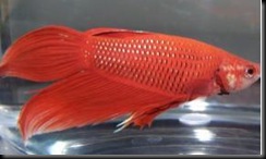
Siamese fighting fish (betta slendens). When Siamese fighting fish encounter certain stressful or threatening environmental conditions, such as exposure to toxic chemicals like mercury, the erythrophores change appearance, and the pigment moves in a characteristic pattern to an internal part of the cell. The change in pigment location in response to a toxic chemical is rapid, obvious and can be numerically described.

This research found that when Siamese fighting fish encounter certain stressful or threatening environmental conditions, such as exposure to toxic chemicals like mercury, the erythrophores change appearance, and the pigment moves in a characteristic pattern to an internal part of the cell. The change in pigment location in response to a toxic chemical is rapid, obvious and can be numerically described.

Another kind of stressful or threatening situation which also causes the location of pigment to change is the toxic threat posed by illness-causing bacteria. Some of these bacteria are associated with food.

"We discovered that the pigment bearing cells, erythrophores, respond immediately to certain food associated, toxin producing bacteria responsible for making humans sick," Trempy said. "There is potential to directly assess the toxic behavior of the contaminating bacteria, not just the simple presence of the DNA or protein of these bacteria. And this response can be easily seen under a low-power microscope and quickly quantified, numerically, to describe the intensity of the situation."

This technology can detect such important food-associated bacteria as Salmonella and Clostridium perfringens, responsible for diarrheal illnesses; Bacillus cereus, responsible for gastrointestinal illness characterized by vomiting and diarrhea, and often referred to as stomach flu, and Clostridium botulinum, which causes toxin-induced botulism, characterized by paralysis.

Further studies are needed to define the pigment bearing cell response to other important bacteria of concern, such as E. coli O157:H7 and Listeria, Trempy said. Research is also needed to immortalize a pigment bearing cell line for mass production and commercial use. These advances should be possible and progress is being made on both issues in continuing research, she said.

It's possible, Trempy said, that portable kits could be developed that would not require specialized training to use. Results would be available in minutes, convenient and would allow food processors, distributors, handlers, or even consumers to quickly assess food for contaminating bacterial toxicity.

Source:ScienceDaily
Wednesday, October 22, 2008

Prebiotic Potential Of Almonds

Almonds, as well as being high in vitamin E and other minerals, are also thought to have other health benefits, such as reducing cholesterol. Recently published work by the Institute of Food Research has identified potential prebiotic properties of almonds that could help improve our digestive health by increasing levels of beneficial gut bacteria.

almonds

Our digestive system maintains large population of bacteria that live in the colon. Prebiotics are non-digestible parts of foods that these bacteria can use to fuel their growth and activity. These 'good' bacteria form part of our body's defence against harmful bacteria and play a role in the development of body's immune system. The prebiotics work by stimulating the growth of these bacteria. However, in order to get to where they are needed prebiotics must be able to get through the upper part of the intestine without being digested or absorbed by the body.

Funded by the Almond Board of California, IFR scientists first used the Model Gut, a physical and biochemical simulator of the gastro-intestinal tract, to subject almonds to the same conditions experienced in the stomach and small intestine. They then added the digested almonds to an in vitro batch system to mimic the bacterial fermentation in the large intestine and monitored its effect on the populations of intestinal bacteria.

The study, published in Applied and Environmental Microbiology, found that finely ground almonds significantly increased the levels of certain beneficial gut bacteria. This effect was not seen when the fat content was removed from the almond preparation, suggesting that the beneficial bacteria use the almond lipid for growth, and this is the basis for the prebiotic effect of almonds.

Previous studies have shown that the amount of available lipid is reduced if the almonds are not processed, for example by grinding as in this study or by chewing. The length of time the almond spends in the digestive system also affects the amount of available lipids and proteins. More detailed studies on the digestibility of almonds are now required, and the prebiotic effect of almond lipids needs to be tested in human volunteers.

Source: ScienceDaily

Tuesday, October 21, 2008

Pillows: A Hot Bed Of Fungal Spores

Researchers at The University of Manchester funded by the Fungal Research Trust have discovered millions of fungal spores right under our noses -- in our pillows.

Aspergillus fumigatus, the species most commonly found in the pillows, is most likely to cause disease; and the resulting condition Aspergillosis has become the leading infectious cause of death in leukaemia and bone marrow transplant patients. Fungi also exacerbate asthma in adults.

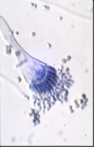
fungal spore

The researchers dissected both feather and synthetic samples and identified several thousand spores of fungus per gram of used pillow - more than a million spores per pillow.

Fungal contamination of bedding was first studied in 1936, but there have been no reports in the last seventy years. For this new study, which was published online today in the scientific journal Allergy, the team studied samples from ten pillows with between 1.5 and 20 years of regular use.

Each pillow was found to contain a substantial fungal load, with four to 16 different species being identified per sample and even higher numbers found in synthetic pillows. The microscopic fungus Aspergillus fumigatus was particularly evident in synthetic pillows, and fungi as diverse as bread and vine moulds and those usually found on damp walls and in showers were also found.

Professor Ashley Woodcock who led the research said: "We know that pillows are inhabited by the house dust mite which eats fungi, and one theory is that the fungi are in turn using the house dust mites' faeces as a major source of nitrogen and nutrition (along with human skin scales). There could therefore be a 'miniature ecosystem' at work inside our pillows."

Aspergillus is a very common fungus, carried in the air as well as being found in cellars, household plant pots, compost, computers and ground pepper and spices. Invasive Aspergillosis occurs mainly in the lungs and sinuses, although it can spread to other organs such as the brain, and is becoming increasingly common across other patient groups. It is very difficult to treat, and as many as 1 in 25 patients who die in modern European teaching hospitals have the disease.

Immuno-compromised patients such as transplantation, AIDS and steroid treatment patients are also frequently affected with life-threatening Aspergillus pneumonia and sinusitis. Fortunately, hospital pillows have plastic covers and so are unlikely to cause problems, but patients being discharged home - where pillows may be old and fungus-infected - could be at risk of infection.

Aspergillus can also worsen asthma, particularly in adults who have had asthma for many years, and cause allergic sinusitis in patients with allergic tendencies. Constant exposure to fungus in bed could be problematic. It can also get into the lung cavities created by tuberculosis which affects a third of the world's population, causing general ill-health and bleeding in the lung, as well as causing a range of plant and animal diseases.

Dr Geoffrey Scott, Chairman of the Fungal Research Trust which funded the study, said: "These new findings are potentially of major significance to people with allergic diseases of the lungs and damaged immune systems - especially those being sent home from hospital."

Professor Ashley Woodcock added: "Since patients spend a third of their life sleeping and breathing close to a potentially large and varied source of fungi, these findings certainly have important implications for patients with respiratory disease - especially asthma and sinusitis."

Source:ScienceDaily

Wednesday, October 15, 2008

'Friendly' Bacteria Protect Against Type 1 Diabetes

In a dramatic illustration of the potential for microbes to prevent disease, researchers at Yale University and the University of Chicago showed that mice exposed to common stomach bacteria were protected against the development of Type I diabetes.

The findings, reported in the journal Nature, support the so-called "hygiene hypothesis" – the theory that a lack of exposure to parasites, bacteria and viruses in the developed world may lead to increased risk of diseases like allergies, asthma, and other disorders of the immune system.

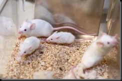
type I diabetes

 Researchers have found that non-obese diabetic mice deficient in innate immunity were protected from diabetes in normal conditions. However, if they were raised in a germ-free environment, lacking "friendly" gut bacteria, the mice developed severe diabetes.

The results also suggest that exposure to some forms of bacteria might actually help prevent onset of Type I diabetes, an autoimmune disease in which the patient's immune system launches an attack on cells in the pancreas that produce insulin. The root causes of autoimmune disease have been the subject of intensive investigation by scientists around the world.

In the past decade, it has become evident that the environment plays a role in the development of some overly robust immune system responses. For instance, people in less-developed parts of the world have a low rate of allergy, but when they move to developed countries the rate increases dramatically. Scientists have also noted the same phenomenon in their labs.

Non-obese diabetic (NOD) mice develop the disease at different rates after natural breeding, depending upon the environment where they are kept. Previous research has shown that NOD mice exposed to killed (i.e., non-active) strains of tuberculosis or other disease-causing bacteria are protected against the development of Type I diabetes. This suggests that the rapid "innate" immune response that normally protects us from infections can influence the onset of Type 1 diabetes.

In the Nature paper, teams led by Li Wen at Yale and Alexander V. Chervonsky at the University of Chicago showed that NOD mice deficient in innate immunity were protected from diabetes in normal conditions. However, if they were raised in a germ-free environment, lacking "friendly" gut bacteria, the mice developed severe diabetes. NOD mice exposed to harmless bacteria normally found in the human intestine were significantly less likely to develop diabetes, they reported.

"Understanding how gut bacteria work on the immune system to influence whether diabetes and other autoimmune diseases occurs is very important," Li said. "This understanding may allow us to design ways to target the immune system through altering the balance of friendly gut bacteria and protect against diabetes."

Source: Sciencedaily.

Friday, October 10, 2008

Toenail Fungus and Athlete’s Foot Do Spread From Person to Person

Cutting-edge molecular biology techniques confirm the long-held but never proven assumption that toenail fungus and athlete’s foot can spread and that the fungus can infect family members living in the same household. Findings have important implications regarding the effective and timely treatment of these infections to prevent their spread throughout the family.

A collaboration between experts in dermatology, epidemiology, and mycology, the study was conducted at five centers led by Dr. Mahmoud Ghannoum, Professor of Dermatology at University Hospitals of Cleveland. The study was supported with funding from Novartis Pharmaceuticals.

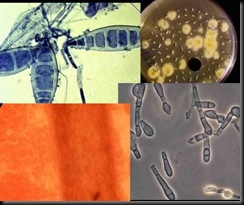
dermaphytes

Affecting more than 35 million people, onychomycosis is a common, chronic, and highly resistant fungal infection in which affected nails become discolored, brittle, thickened, and flaky. Onychomycosis often results from tinea pedis, another common fungal skin infection affecting 10% of the general population at any time. Both infections are caused by tiny organisms called dermatophytes; the most common dermatophytes are Trichophyton rubrum (T. rubrum), Trichophyton mentagrophytes (T.mentagrophytes), and Epidermophyton floccosum (E. floccosum).

While it has long been assumed that dermatophytes spread from person to person, there has never been medical or scientific evidence supporting this contention. By providing scientific confirmation that dermatophytes can be transmitted among family members, this study suggests that toenail fungus and tinea pedis are infections that should be treated aggressively to help prevent their spread. 

The prospective study enrolled 57 families in which at least one member, the “index person,” had either nail fungus and/or athlete’s foot. Of these, 19 families were identified in which at least two members were infected.  Using conventional and culture growth methods to examine a total 110 toenail and skin samples taken from every participant, researchers determined that the vast majority of samples (80.4 %) were of the species T. rubrum, while most of the others were infected with T. mentagrophytes (14.3%), and E. floccosum. The identity of each species was confirmed using polymerase chain reaction (PCR), a molecular technique in which a species-specific fragment of DNA is amplified millions of times. Using a more sophisticated molecular biology technique called restriction fragment length polymorphism (RFLP), these amplified DNA molecules are further analyzed. Combining these two techniques, researchers were able to determine the identity of each strain based on unique DNA band patterns.

Results showed transmission of identical dermatophytes among 42% (8/19) of families. Among the families where spread of infection among members was detected, 87.5% (7/8) were infected with T. rubrum. Analysis of DNA band patterns using RFLP revealed that 62% (5/8) of the families were infected with the same strain of T. rubrum (Type D). Matching the DNA of dermatophytes among members of the same household clearly indicated that each family member had the identical strain of dermatophyte, and confirmed that the infection had been transmitted from one family member to the other  either directly or indirectly.

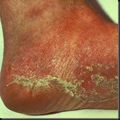
Tinea pedis

In addition, the results of a questionnaire answered by the index person and family members identified the following as factors that significantly correlated with spread of infection in a household: past history of onychomycosis and tinea pedis, having tinea pedis for more than 10 years, and the occurrence of tinea pedis with scaling of the skin on the side of the foot.

The novel findings of this research suggest areas for future study, looking at factors that might determine why some family members became infected while others did not, as well as why the infection spread only within some of the families. Whether some strains may be more virulent, and if some individuals may be genetically susceptible to fungal infection, are other questions this study raises for future investigation

Source: American Society for Microbiology

Monday, October 6, 2008

Bacteria ready to make plastic from sugar

Discovery may replace petroleum-based ingredients with renewable ones

Claims of biology-based oil and plastic usually bear the caveat "in five years." But a San Diego-based company claims they will have a pilot plant for production of E.coli-based 1,4 butanediol (BDO), the base chemical for plastic products ranging from Spandex to car bumpers, next year.

"We are able to couple the growth of the organism to the production of BDO," said Christophe Schilling, co-founder of the company, Genomatica. "For the bacteria to grow they have to produce BDO."

The announcement holds particular promise amid rising oil prices and a scramble to replace petroleum-based ingredients with renewable ones. Traditionally, BDO is made from oil and natural gas through an energy-intensive process. Genomatica, founded in 2000, says their technology can significantly reduce the cost of making BDO. All they need is sugar and a particular bacterium.

The specific cost saving will vary by company, but Chris Gann, Genomatica's CEO, says that, "if the price of oil dropped to $50 per barrel [our technology] would still be competitive." The price for a barrel of oil earlier this week was about $120.

That savings in cost comes from using cheaper raw materials and less energy to trigger the chemical reactions necessary to turn oil into plastic, Gann said.

Using computer-aided design (CAD) software similar to what engineers have used for years, Genomatica scientists simulated various chemical reactions to find the best genes for the job.

Schilling won't say from which specific organisms they got the proteins and enzymes used to turn sugar into BDO, other than that they culled them from a computer database of "hundreds of microorganisms," some obscure, some commonplace.

Once Genomatica scientists had a working virtual E. coli, they spliced the selected genes into the real E. coli and turned them loose on raw cane sugar.

E.coli

So far, Genomatica has produced less than two pounds of BDO. To create industry-scale amounts of BDO, cane sugar, E. coli and water will go into 105-degree (Fahrenheit) fermentation tanks, like the ones used to produce ethanol.

Still, cells are notorious for evolving unwanted traits, like the rise of drug resistant bacteria and cancers. Schilling said they plan to tap into that evolutionary bent and turn it into profit.

"We have ways to essentially accelerate [evolution], so the bacteria can evolve to tolerate higher concentrations of BDO and not in the exact way we would have predicted with the computer models," said Schilling.

Genomatica will make the E. coli, but not at the plants where the BDO is produced. Instead, the company will license the genetically engineered E. coli to companies worldwide who create BDO-based products. The company can even custom design E. coli that take advantage of local sugar variations. The first pilot plant is set for next year, according to Genomatica.

Next year might be a bit ambitious, suggested Harvey Blanch, a professor of chemical engineering at the University of California, Berkeley. Blanch said a longer time line, as in a couple of years, was more likely, but that he was still quite excited by the development.

"This is a very interesting opportunity to get a new source of raw materials that could replace chemical processing with biological engineering," said Blanch. "Using this technique everything from new kinds of plastics to jet fuel could be produced with less energy and smaller environmental costs."

Genomatica hopes that bio-BDO is just the first of these new raw materials. Currently, the company is pursuing more than six other unspecified chemicals, all of which have a larger market than the $4 billion annual BDO market."What gets us really excited is that this opens up the door to go after other chemicals that aren't produced in nature," said Schilling

Sources: Msnbc

Thursday, October 2, 2008

Tinkering extends life of organism by 10-fold

Genetic breakthrough could ultimately inform efforts on humans

Scientists have extended the lifespan of yeast, microbes responsible for creating bread and beer, by 10-fold. That's twice the previous record for life extension in an organism. The breakthrough could ultimately inform efforts to extend human lives. Instead of one week, the yeast lived for about 10 weeks through genetic tinkering and a low-calorie diet. Brewer's yeast, known formally as Saccharomyces cerevisiae, illuminated using a special technique called immunofluorescence. Scientists have recently extended the lifespan of the microscopic organism by both tinkering with aging genes and cutting the amount of calories it takes in

"We've reprogrammed the healthy life of an organism," said Valter Longo, a biologist at the University of Southern California in Los Angeles who led the life-prolonging experiments.

 Genetic soldiers
DNA, short for deoxyribonucleic acid, is the body's set of blueprints and instructions, carried by genes.
"Evolution designed our genes, our army, to be ready for growth and reproduction," Longo told LiveScience. Problem is, pooling the body's efforts into growing makes room for genetic errors that lead to age-related disease. "We can use our energy to grow and reproduce, or protect ourselves."

Longo and his team previously found two genes — RAS2 and SCH9 — related to growth and development of cancer that are similar in humans and yeast. They are so alike, in fact, that Longo said, "you can put the human gene in yeast and it works."

The scientists disabled the genes in the yeast but also put the organism on a low-calorie diet. Caloric restriction has prolonged the lifespan of yeast, worms, and mice in other experiments, and is thought to work by scaring the body into maintaining its genetic goods instead of growing.

Combining both age-fighting approaches, Longo said, led to a dramatically long lease on life. "We expected a small boost in longevity, but not a 10-fold increase," he said. "It's remarkable." Longo thinks the genes act like generals of the genetic army, ordering the troops to protect the body's DNA under caloric stress instead of fighting for growth.“I would say 10-fold is pretty significant,” said Anna McCormick, chief of the genetics and cell biology branch at the National Institute on Aging in Bethesda, Md., of Longo's findings.

Hope for humans?

To find out how the age-defying treatment works in humans, Longo and his group are now studying Ecuadorians who have similar mutations in age-controlling genes used in the yeast. “People with two copies of the mutations have very small stature and other defects,” he said. Despite the problems, Longo said, the people likely benefit from their condition.

"So far, we have never seen cancer in people who have two copies of the mutated genes," he said. “We are now identifying the relatives with only one copy of the mutation, who are apparently normal. We hope that they will show a reduced incidence of diseases and an extended life span.”

Longo thinks life-extending drugs that have no major side effects will not be easy to develop but should be possible in the future. He explained that manipulating the genes leads to major growth defects probably because they are inactive during childhood.

"What if we could achieve a balance by switching those genes off when we want to?" he asked. "Twenty or thirty years from now, we might have the ability to reduce the activity of [the genes]. In the long run, I think that balance may not be too hard to achieve.

Sources: Msnbc